
Otitis externa in small animals
The structure of the ear
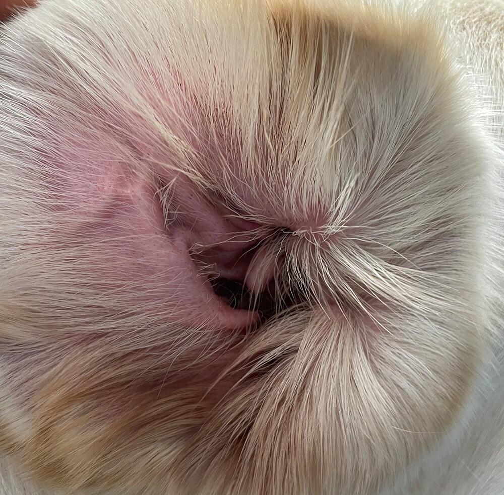
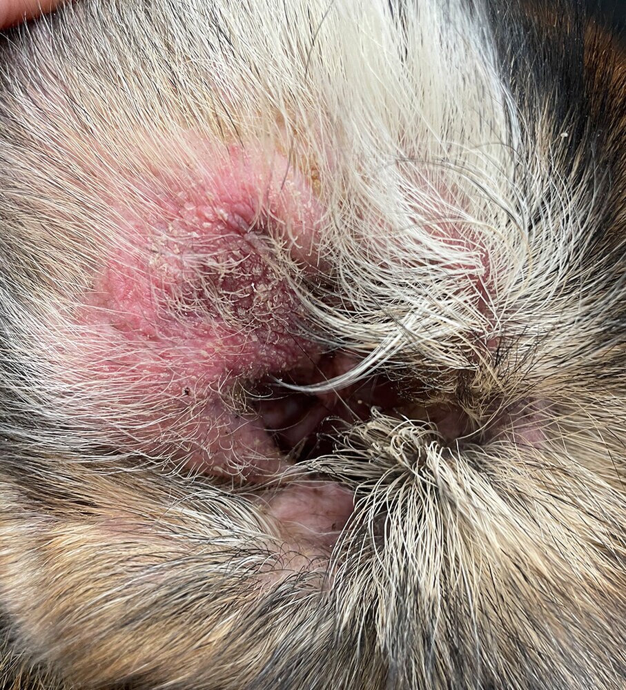
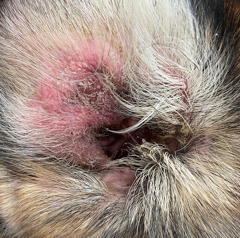
The ear consists of the outer, middle and inner ear. The outer auditory canal ends with the eardrum. Like the rest of the body, it is lined with epithelium, hair and skin appendage glands. When we talk about otitis externa, we mean an inflammation in the area of the pinna and the external auditory canal (Fig. 1, Fig. 2).
Why do ear infections occur?
There are many causes of ear infections. As with all skin problems, a precise medical history is important for successful treatment.
A distinction is made between various factors that influence the development of an ear infection:
- Predisposing factors: narrow or very hairy ear canals, for example, favour the development of an inflammation. However, increased earwax production or frequent swimming can also lead to otitis externa.
- Primary causes of the disease: These are common and range from foreign bodies, parasitic infections and allergies to hypothyroidism, polyps and tumours through to autoimmune diseases such as pemphigus foliaceus.
- Perpetuating factors can maintain or exacerbate an ear infection: resistant infections, otitis media, changes in the ear canal (swelling, scarring or ossification causing narrowing of the ear canal).
What does inflammation of the ear canal mean?
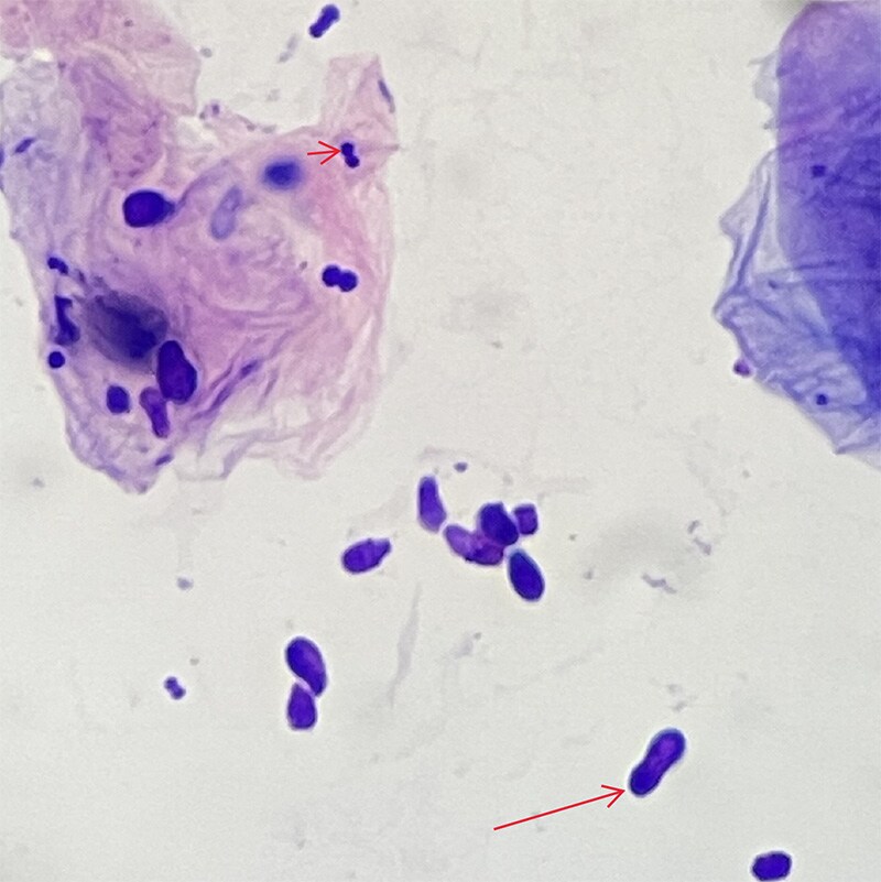
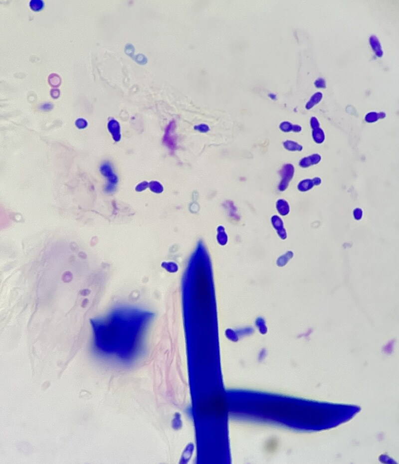
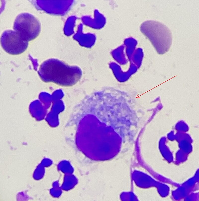
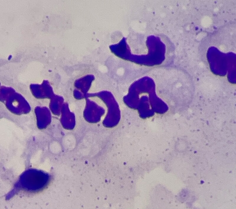
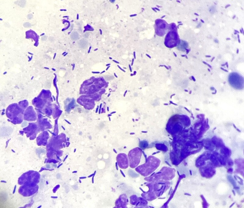
A colonisation of the external auditory canal with certain germs is normal; every healthy ear has a distinct natural mixed flora. However, if there is an inflammation, certain pathogens are usually present in excess. A distinction is made between bacterial and/or yeast overgrowth (Fig. 3, Fig. 4) or infection (detection of bacteria and/or malassezia). An overgrowth occurs as soon as more bacteria or yeasts are detected than normal. An infection is present if inflammatory cells (Fig. 5, Fig. 6) are also found or certain pathogenic bacteria (Fig. 7a, b, Fig. 8) are present.
How do you examine the external auditory canal?
Animals with ear infections show different symptoms: Some have itching or pain in the ear area, others show head shaking or tilting. In addition, an increased amount of cerumen (earwax) or purulent (sometimes smelly) material may be visible.
Examination of the pinna shows whether other skin changes are present. By palpating the head, you can feel swellings or indurations and evaluate whether itching or pain is present. The ear canal itself is examined using an otoscope lamp. This is the only way to assess the horizontal area of the ear canal and the eardrum. Space-occupying processes, the condition of the cerumen/exudate, foreign bodies and the presence of inflammatory symptoms are assessed in this way.
Tip:
Have two otoscope attachments ready. It is important to use a separate attachment for each ear so as not to transfer possible different infections from one ear to the other.
In some cases, otoscopy is not possible straight away, e.g. if the patient is too painful or the ear canal is very swollen. In these cases, sedation is sometimes necessary for the examination or decongestant therapy must first be administered to enable the otoscopy to be performed at a later date.
Good to know
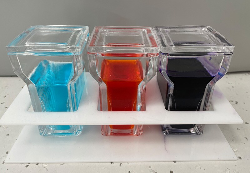
This is how easy a quick stain is: For the cytological examination (Fig. 9), the slide is stained with a Diff-Quick stain (Fig. 10), which is typically used in practice to assess blood smears and cytological samples. This involves a fixing solution and two staining solutions.
After drying the smear (air drying or brief heat fixing), the specimen is immersed in each solution for 5 seconds: first in the fixing solution, then in the red and finally in the blue staining solution. The slide is then carefully rinsed with distilled water. After drying, the sample can be examined under a microscope: This can determine whether there is an overgrowth/infection with bacteria and/or yeast fungi or inflammatory cells, which gives a decisive indication of the treatment to be carried out or further diagnostics. A small number of cocci (round bacteria) and malassezia (yeast fungi) should be considered normal, whereas the presence of rod-shaped bacteria and inflammatory cells should always be considered pathological.
This examination technique is also ideal for evaluating the success of treatment, as the quantity of bacteria present can be easily assessed in the impression.
What role do microscopy and cytology play?
The type of inflammation/infection present is diagnosed using a very simple examination method: A swab is taken from the ear canal and examined under a microscope (Fig. 9). This examination only takes a few minutes and can be carried out on practically every patient.
Materials required:
- Cotton swab
- Microscope slide
- Haema rapid staining (Diff-Quick, Fig. 10)
- Microscope
Using a cotton bud, collect a sample from the transition from the vertical to the horizontal auditory canal with a rotating movement. The cotton swab is then rolled out onto a microscope slide.
Tip:
If you suspect a parasitic infection, you can examine the slide directly, i.e. natively, under the microscope at the lowest magnification.
When is a bacteriological culture and resistance test useful?
By taking sterile swab samples of tissue, bacteria can be determined in the laboratory with subsequent resistance testing. This allows the exact bacterial strain to be diagnosed and provides information regarding the possible development of resistance of the germs in relation to antibiotic therapy.
This examination method should be used if:
- recurrent ear infections are present
- repeated courses of antibiotics have not been successful
- an inflammation of the middle ear is suspected
- rod-shaped bacteria are found in the cytological examination
As there has been an increase in infections with multi-resistant germs in recent years and this issue also affects veterinary medicine, the use of antibiotics should be precisely indicated and the success of treatment should be carefully monitored.
Successful treatment of otitis externa
The successful treatment of ear infections depends on various factors: In addition to the correct treatment of otitis externa, the underlying factors must not be neglected. Further measures must be carried out in a targeted manner in order to diagnose or treat primary problems. Otherwise, the risk of recurring inflammation is very high.
Tip:
Owner communication is very important: by conveying a good understanding of the causes and necessary therapeutic measures, you make a decisive contribution to successful treatment.
The right expertise for successful therapy
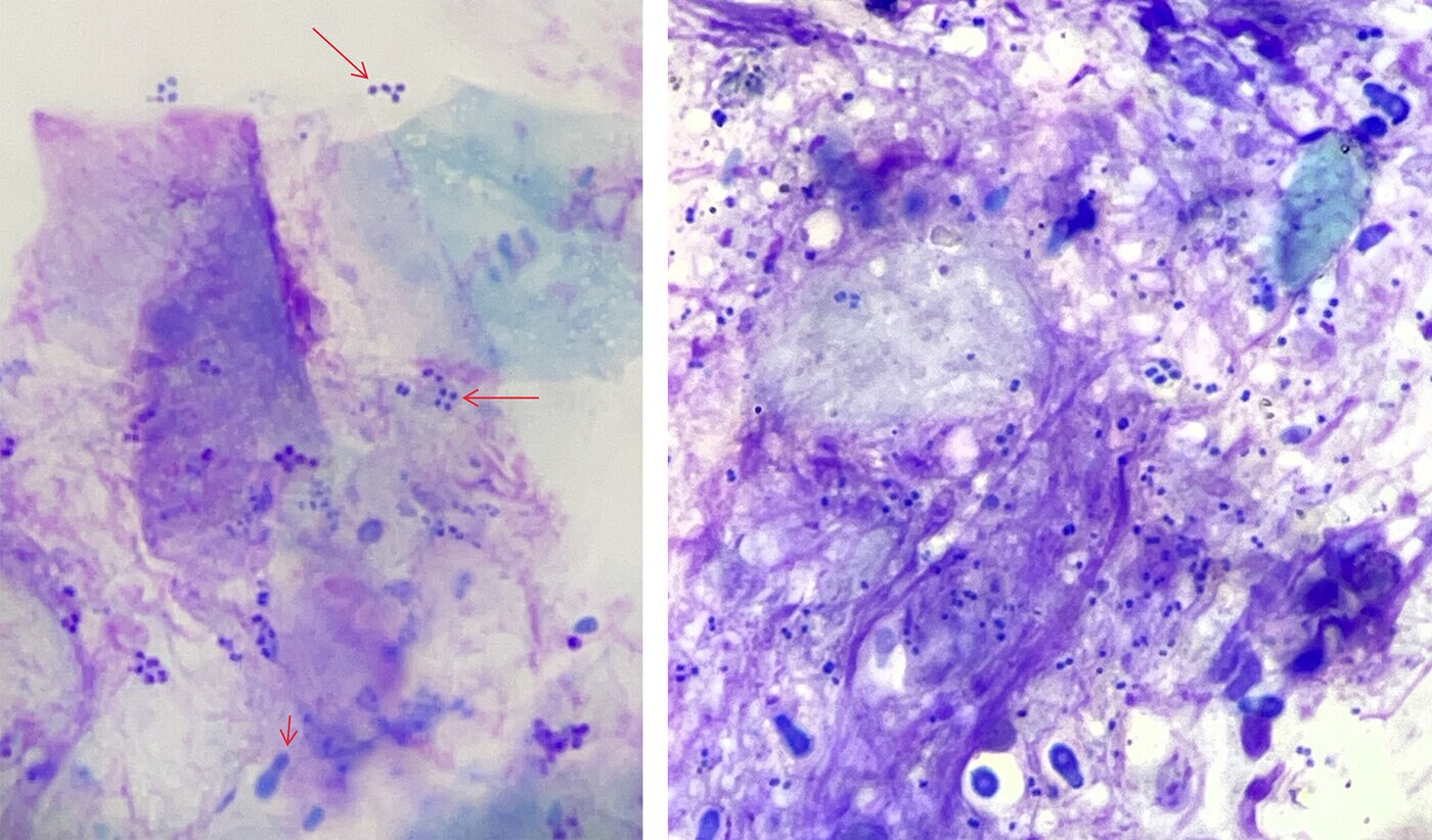
We have various means at our disposal for the treatment of otitis externa. Mechanical removal of cerumen and infectious material is particularly important as part of the therapy. The frequency of use and choice of cleanser depend on the severity and type of inflammation. Bacteria can form a protective matrix around themselves (biofilm, Fig. 11). This makes them insensitive to antibiotics and certain environmental influences. Good cleaning of the ear canal is particularly important in these cases.
Ear cleaners and active ingredients:
- Zerumenolytic agents help with firmly adhering plaque and dissolve it well after a short exposure time.
- If the ear canal is very moist, drying agents help.
- The more crumbly and crusty the lesions are, the more suitable oily cleaners are for treatment.
- If the ear canal is more weeping and moist, aqueous substances help.
- Antimicrobial ingredients have a good effect on bacterially infected otitis and are used alone or in combination with antibiotic substances. They are an important therapeutic agent for chronic and multi-resistant infections.
In general, local therapy is preferable to systemic treatment for otitis externa. The choice of suitable medication depends on the type of infection. Some active substances are only intended for the external auditory canal (potentially ototoxic), which is why otoscopic examination of the auditory canal and assessment of the eardrum are important for choosing the appropriate medication.
Short and sweet
Otoscopy and cytology are essential for a correct examination of otitis externa. The cytological examination provides quick and easy information about the type and severity of the inflammation and helps in the selection of further diagnostic and therapeutic measures. In order to successfully treat otitis externa, underlying causes must also be treated. Good communication with the patient's owners and precise training on the correct cleaning/treatment of the ear are crucial.
All images © Dr Barbara Heilinger

Dr Barbara Heilinger
Veterinary dermatologist
https://vetdermatologie.at
Former head of the dermatological outpatient clinic at the University of Veterinary Medicine, Vienna